






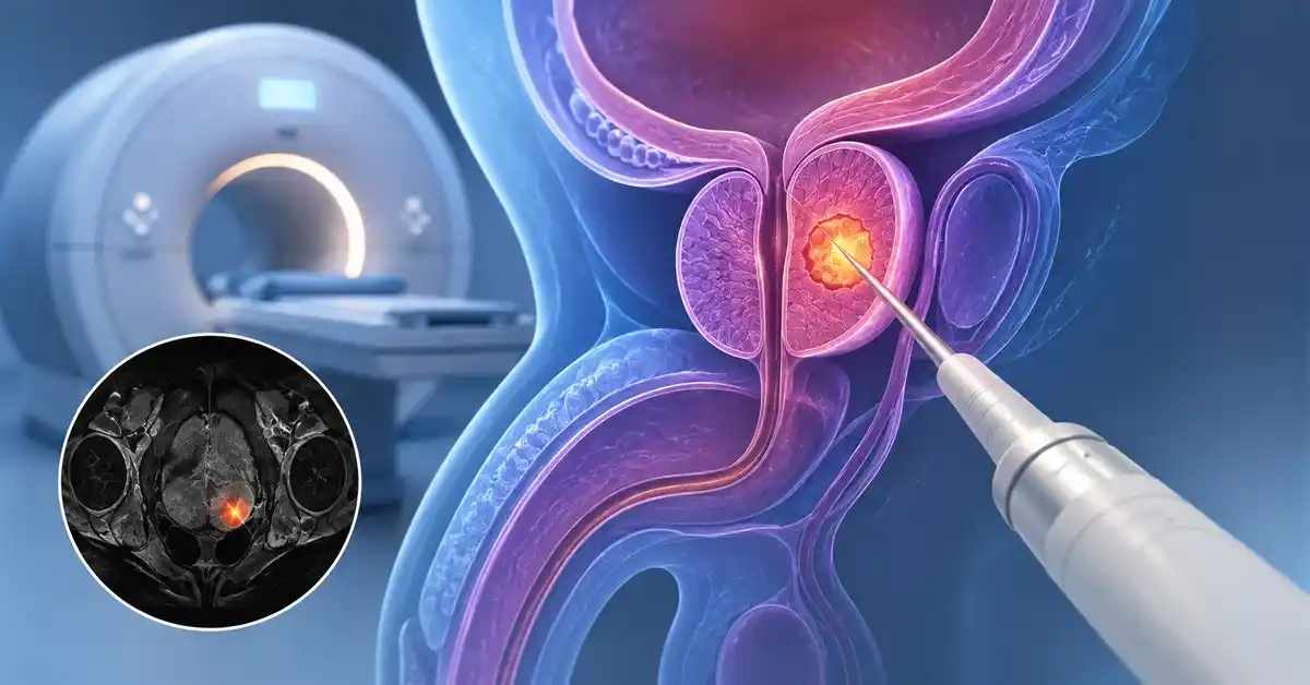
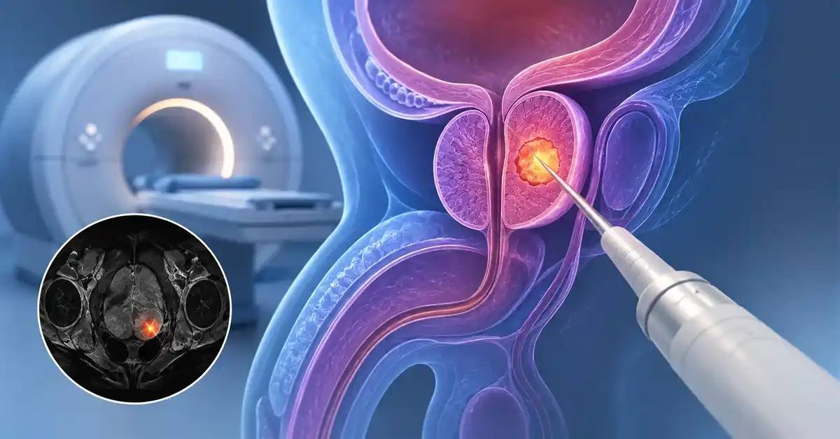
Multiparametric prostate MRI has significantly changed the diagnostic pathway for suspected prostate cancer. Instead of proceeding immediately from an elevated PSA to relatively uniform sampling across the gland, MRI can identify suspicious lesions that deserve focused biopsy.
The commonly used phrase “MRI-guided prostate biopsy” usually refers to MRI-targeted biopsy. The MRI is obtained first, suspicious areas are mapped, and tissue samples are then directed toward these targets using MRI-ultrasound fusion, cognitive targeting, or, in selected cases, direct in-bore MRI guidance.
The key advantage is greater precision in sampling areas more likely to contain clinically significant cancer. However, targeted biopsy does not guarantee that every tumor in the prostate will be detected, and additional regional or systematic cores may still be appropriate according to the patient’s overall risk.
How Prostate MRI Supports Earlier Cancer Detection
Multiparametric MRI provides anatomical and functional information about different regions of the prostate and can identify lesions that look different from surrounding tissue.
It should nevertheless be interpreted together with PSA, prostate volume, examination findings, age, family history, and previous biopsy information rather than used as a stand-alone cancer test.
Why Is MRI Performed Before Prostate Biopsy?
MRI may be requested because of:
- Persistently elevated PSA.
- An abnormal prostate examination.
- High PSA density.
- Strong family history.
- Ongoing suspicion after a previous negative biopsy.
- Need to locate a lesion before repeat biopsy.
- Risk assessment before surveillance or treatment decisions.
The scan can help identify areas that conventional sampling might otherwise miss, including some anterior lesions.
What Does a PI-RADS Score Mean?
Suspicious lesions are commonly categorized from 1 to 5 according to the likelihood that clinically significant prostate cancer may be present.
| Score | General interpretation |
|---|---|
| PI-RADS 1 | Very low suspicion |
| PI-RADS 2 | Low suspicion |
| PI-RADS 3 | Indeterminate |
| PI-RADS 4 | High suspicion |
| PI-RADS 5 | Very high suspicion |
A PI-RADS 4 or 5 lesion does not prove cancer, while a low score does not exclude it completely.
Does a Negative MRI Mean Biopsy Is Unnecessary?
Not in every patient.
A negative MRI can help some lower-risk men avoid immediate biopsy and instead undergo structured follow-up. Biopsy may still be considered when other factors remain concerning, including:
- High PSA density.
- Abnormal clinical examination.
- Strong family history.
- Important inherited risk.
- Persistently concerning PSA behavior.
- High overall clinical suspicion.
MRI therefore modifies risk rather than reducing the decision to a simple positive-or-negative result.
Read about: Prostate Enlargement Treatments: Medication or Surgery
How MRI-Targeted Prostate Biopsy Is Performed
MRI identifies the target, but another method is needed to guide the needle toward it. Several targeting techniques are available.
The biopsy can also be performed through the perineum or through the rectum.
MRI-Ultrasound Fusion Biopsy
Fusion biopsy electronically combines the previously acquired MRI with live ultrasound imaging during the procedure.
The process generally involves:
- Identifying the lesion on MRI.
- Imaging the prostate with ultrasound.
- Aligning both image sets.
- Navigating the biopsy needle to the target.
- Obtaining several tissue cores from different parts of the lesion.
This method allows MRI planning to be used during real-time biopsy.
Cognitive Targeting and In-Bore MRI Biopsy
With cognitive targeting, the physician reviews the MRI and mentally maps the lesion to the corresponding area seen on ultrasound.
In-bore biopsy is performed while the patient is positioned within an MRI system, allowing direct confirmation of needle placement. It requires specialized equipment and is not necessary for most patients.
The best approach is the one that provides reliable targeting for the individual lesion and clinical setting.
Transperineal Versus Transrectal Biopsy
| Feature | Transperineal | Transrectal |
|---|---|---|
| Needle path | Through perineal skin | Through rectal wall |
| Passage through bowel | No | Yes |
| Infection risk | Generally lower | Relatively higher |
| Access to anterior prostate | Excellent | Possible but sometimes less direct |
| Rectal bleeding | Not expected from needle route | Can occur |
| Urinary retention | Possible | Also possible |
| Anesthesia | Depends on technique | Depends on technique |
The transperineal approach has become increasingly attractive because of its infection profile and ability to sample the full gland.
When Is Biopsy Needed After Elevated PSA or an MRI Lesion?
An elevated PSA does not automatically mean prostate cancer.
PSA may increase with benign enlargement, inflammation, urinary infection, recent instrumentation, and other factors. The biopsy decision should therefore reflect overall risk rather than one blood-test result.
Interpreting PSA Before Biopsy
Assessment can include:
- Current PSA.
- Previous PSA values.
- Prostate volume.
- PSA density.
- Age.
- Family history.
- Examination findings.
- MRI findings.
- Recent factors capable of altering PSA.
PSA density relates the PSA value to the size of the prostate and can provide additional context when interpreting an elevated result.
When Does an MRI Lesion Become More Concerning?
Concern generally rises when a higher-suspicion lesion occurs together with additional clinical risk factors.
Indeterminate lesions require more individualized interpretation. PSA density, lesion size, previous biopsies, age, family history, and overall cancer probability can all influence whether tissue sampling is recommended.
What If a Previous Biopsy Was Negative?
Persistent suspicion may justify further investigation.
One benefit of MRI is that it can reveal a suspicious area that may have been poorly sampled during a previous systematic biopsy.
A repeat procedure can then specifically target that lesion rather than simply repeating the previous sampling pattern.
Read about: Latest Non-Surgical Treatments for Enlarged Prostate
MRI-Targeted Versus Systematic Prostate Biopsy
Targeted biopsy samples the visible MRI lesion, while systematic sampling obtains tissue from predefined or regional areas of the gland.
The two approaches provide partly different information.
Advantages of Targeted Biopsy
Targeted cores can help:
- Focus sampling on higher-risk lesions.
- Improve characterization of MRI-visible abnormalities.
- Relate pathology to a known anatomical target.
- Detect clinically important disease efficiently.
- Reduce unnecessary sampling in selected strategies.
The technique is particularly valuable when a clear lesion is present.
Why Additional Cores May Still Be Taken
MRI is not perfect, and targeting is not mathematically exact.
A significant cancer focus can occasionally exist away from the visible target, and targeted cores can sometimes underestimate tumor grade. Current evidence therefore supports complementary sampling in appropriate patients.
The diagnostic plan attempts to balance adequate cancer detection against unnecessary detection of very low-risk disease.
Which Biopsy Strategy Is Best?
| Clinical situation | Possible approach |
|---|---|
| Clear MRI lesion, first biopsy | Targeted plus additional regional sampling as appropriate |
| Previous negative biopsy with new target | Strong emphasis on targeted sampling |
| Negative MRI with low overall risk | Surveillance may be considered |
| Negative MRI with high clinical risk | Biopsy may still be appropriate |
| Multiple MRI lesions | Individual targeting of relevant lesions |
| Anterior lesion | Transperineal access can be particularly useful |
The objective is sufficient diagnostic information rather than simply maximizing the number of cores.
Prostate Biopsy Procedure, Recovery, and Possible Side Effects
The biopsy itself is usually relatively short, although preparation varies according to the route, anesthesia, medical history, and medication use.
Anticoagulant and antiplatelet medications should be reviewed before the procedure rather than stopped or continued without specific instructions.
Main Steps of MRI-Targeted Biopsy
The procedure may include:
- Reviewing MRI targets.
- Reviewing medications and bleeding risk.
- Checking for urinary infection when indicated.
- Positioning the patient.
- Administering local or planned anesthesia.
- Imaging the prostate with ultrasound.
- Aligning MRI and ultrasound if fusion technology is used.
- Sampling each suspicious target.
- Obtaining additional regional cores when appropriate.
- Labeling specimens by location for pathological examination.
Accurate specimen labeling allows the pathology result to be matched to the MRI lesion.
What Is Normal After Biopsy?
Many patients can return home the same day.
Temporary effects may include:
- Mild blood in the urine.
- Blood in semen, which can remain visible longer.
- Local discomfort.
- Mild bruising after transperineal biopsy.
- Temporary difficulty urinating.
Patients should follow their specific instructions regarding activity, fluids, medications, and follow-up.
When Is Urgent Medical Review Needed?
Seek prompt assessment for:
- Fever or chills.
- Significant deterioration in general condition.
- Inability to urinate.
- Heavy or persistent bleeding.
- Severe increasing pain.
- Fainting or marked dizziness.
- Symptoms suggesting infection.
Transperineal biopsy generally carries a lower infectious risk, although complications remain possible.

Understanding Biopsy Results and Their Role in Early Diagnosis
The purpose of biopsy is not merely to label cancer as present or absent. Tissue analysis also helps determine tumor grade, volume within the cores, and biological characteristics that influence treatment decisions.
This is particularly important because not every prostate cancer identified through early detection requires immediate treatment.
What Does the Pathology Report Show?
A report may describe:
- Whether cancer is present.
- Histological type.
- Grade Group.
- Tumor pattern.
- Number of positive cores.
- Amount of cancer in individual cores.
- Additional important pathological features.
These findings are then interpreted alongside PSA and imaging.
What Happens With Low-Risk Prostate Cancer?
Some prostate cancers grow slowly and may have a very low probability of affecting health during the foreseeable future.
Active surveillance may therefore be appropriate for selected low-risk patients. Surveillance can include PSA testing, clinical review, MRI, and repeat biopsy when indicated.
Early detection should therefore aim for clinically meaningful diagnosis, not automatic treatment of every small cancer.
What Happens When Clinically Significant Cancer Is Found?
Management depends on:
- Grade.
- Tumor amount.
- Stage.
- PSA.
- Imaging.
- Patient age.
- General health.
- Evidence of disease beyond the prostate.
Treatment options can include surveillance in carefully selected situations, surgery, radiotherapy, systemic approaches, or combinations according to disease risk.
Read about: Interventional Radiology for Enlarged Prostate Treatment
Conclusion
MRI has changed prostate-cancer diagnosis by allowing suspicious areas to be identified before biopsy and directing tissue sampling toward those targets. This improves the ability to investigate clinically important lesions while helping some lower-risk patients avoid unnecessary immediate biopsy.
However, MRI does not replace tissue diagnosis when biopsy is indicated, and targeted biopsy alone is not necessarily sufficient for every patient. PSA, PSA density, MRI findings, family history, previous biopsy results, and overall risk should be interpreted together.
Frequently Asked Questions: MRI-Targeted Prostate Biopsy: Early Diagnosis
Can prostate MRI replace biopsy?
Not always. MRI may allow some lower-risk patients to avoid immediate biopsy, but tissue is required when histological confirmation is necessary.
Does PI-RADS 5 mean cancer is certain?
No. It indicates very high suspicion but remains an imaging assessment rather than a definitive diagnosis.
Is transperineal biopsy preferable to transrectal biopsy?
It offers excellent access to the prostate and generally has a lower infection risk, making it increasingly used.
Can biopsy be negative even when cancer is present?
Yes. A biopsy samples only portions of the gland, so continued follow-up may be needed when suspicion remains high.
How quickly are prostate-biopsy results available?
Timing varies by laboratory and required pathological testing. The result should be interpreted together with PSA and MRI findings.