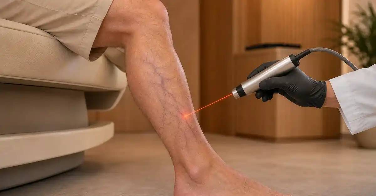








Varicose veins are more than enlarged vessels visible beneath the skin. They can reflect abnormal venous return caused by valves that no longer close effectively, allowing blood to flow backward and raise pressure within superficial leg veins.
Symptoms can begin before large, twisted veins become obvious. Leg heaviness, aching after prolonged standing, and mild ankle swelling at the end of the day can all occur in chronic venous disease. Diagnosis therefore combines symptoms, physical examination, and duplex ultrasound when confirmation or treatment planning is needed.
Early Signs of Varicose Veins Before Large Veins Appear
Venous disease can begin gradually, and symptoms may precede the classic appearance of bulging veins.
Leg Heaviness and Aching
Typical symptoms can include:
- Heavy legs.
- Aching or throbbing.
- Burning or tightness.
- Symptoms that worsen after prolonged standing.
- Partial relief after walking or elevating the legs.
The relationship between symptoms and prolonged standing is useful during clinical evaluation.
Ankle and Lower-Leg Swelling
Mild ankle swelling can develop toward the end of the day and improve after overnight rest.
As venous disease progresses, edema can become more persistent. However, leg swelling also occurs with cardiac, renal, medication-related, lymphatic, and thrombotic conditions, so edema alone does not diagnose varicose veins.
Itching, Cramping, and Local Discomfort
Some people report itching, cramps, pulsation, or discomfort around visible veins.
These symptoms are not specific to venous disease but become more suggestive when combined with enlarged veins, edema, or skin changes.
Varicose Veins Versus Spider and Reticular Veins
Visible superficial veins exist on a spectrum from tiny telangiectasias to large varicosities and advanced chronic venous insufficiency.
Spider and Reticular Veins
Spider veins are small red, blue, or purple vessels near the skin surface.
Reticular veins are larger and often appear blue or green beneath the skin.
They can sometimes be primarily cosmetic, although symptoms or deeper venous abnormalities may coexist. The need for ultrasound depends on the clinical context rather than appearance alone.
What Do True Varicose Veins Look Like?
Varicose veins are typically enlarged, tortuous, and raised above the skin.
They are commonly seen along the calf, behind the knee, or along the inner thigh and lower leg and often become more prominent when standing.
Skin Changes in More Advanced Venous Disease
Progressive venous hypertension can lead to:
- Persistent edema.
- Brown or dark pigmentation.
- Venous eczema.
- Hardening of tissues near the ankle.
- Healed venous ulcers.
- Active venous ulcers.
Clinical classification systems describe this progression from small surface veins through varicosities, edema, skin disease, and ulceration.
Risk Factors for Developing Varicose Veins
Some people develop varicose veins without an obvious trigger, but several factors increase susceptibility.
Family History and Age
A family history of varicose veins or chronic venous disease increases risk.
Venous disorders also become more common with aging as vein walls, valves, and surrounding tissues change.
Pregnancy, Body Weight, and Prolonged Standing
Pregnancy changes venous pressure and blood flow in the lower limbs.
Obesity and increased abdominal pressure can also place additional stress on venous return, while long periods of standing or sitting can aggravate symptoms in susceptible people.
Previous Deep Vein Thrombosis
Previous DVT is particularly important because it can damage deep-vein valves or leave chronic obstruction.
This can produce post-thrombotic venous insufficiency that differs from uncomplicated superficial varicose veins.

When Varicose Veins Should Be Medically Assessed
Small asymptomatic spider veins do not require the same evaluation as painful varicosities with edema or skin damage.
Symptoms That Deserve Venous Assessment
Assessment becomes more important with:
- Recurrent aching or heaviness.
- Persistent or recurrent swelling.
- Large painful varicose veins.
- Itching or dermatitis near the ankle.
- Skin discoloration.
- A wound that heals poorly.
- Recurrent superficial vein inflammation.
- Recurrent veins after previous treatment.
These findings can indicate clinically significant reflux or obstruction.
Leg Pain Is Not Always Venous
Pain can also come from musculoskeletal, neurological, arterial, spinal, or thrombotic disease.
A clinical examination and targeted imaging help determine whether visible veins actually explain the patient’s symptoms.
Warning Signs Requiring Prompt Assessment
Sudden one-sided leg swelling, pain, and warmth require timely assessment because acute venous thrombosis must be excluded.
Heavy bleeding from a superficial vein, an infected ulcer, or new shortness of breath or chest pain in someone with leg symptoms also requires urgent medical evaluation.
How Doctors Diagnose Varicose Veins and Venous Reflux
Diagnosis begins with history and physical examination. Duplex ultrasound is the main imaging test when venous reflux, obstruction, or treatment planning needs to be evaluated.
Physical Examination While Standing
The patient may be examined while standing because gravity makes varicose veins and venous filling more apparent.
The clinician looks for:
- Distribution of visible veins.
- Ankle and leg swelling.
- Asymmetry.
- Skin pigmentation.
- Eczema.
- Healed or active ulcers.
- Signs of previous thrombosis or surgery.
The appearance alone cannot reliably identify the underlying refluxing vein.
Duplex Venous Ultrasound
Duplex imaging combines anatomical ultrasound with Doppler analysis of blood flow.
It can determine:
- Whether the deep veins are open.
- Whether thrombosis is present.
- Whether abnormal backward flow exists.
- Which superficial vein is responsible.
- How extensive the reflux is.
- Whether perforating veins are involved.
Testing for reflux is commonly performed with positional and compression maneuvers designed to challenge venous valve function.
When Are CT or MR Venography Needed?
Most uncomplicated varicose veins do not require advanced cross-sectional imaging.
CT or MR venography may be considered in selected complex cases when pelvic or abdominal venous obstruction is suspected or when symptoms cannot be adequately explained by routine ultrasound.
How Diagnosis Determines the Severity and Treatment Plan
The purpose of evaluation is not simply to confirm that an enlarged vein exists. It is to identify the origin of reflux and determine whether the disease has begun to affect the skin and surrounding tissues.
Clinical Stages of Venous Disease
| Clinical stage | Typical finding |
|---|---|
| Spider or reticular veins | Small superficial vessels |
| Varicose veins | Enlarged tortuous veins |
| Edema | Persistent swelling |
| Skin changes | Pigmentation, eczema, or fibrosis |
| Healed ulcer | Previous venous wound |
| Active ulcer | Open venous wound |
Clinical staging supports consistent description, while duplex ultrasound defines the underlying venous anatomy and hemodynamics.
Why the Source of Reflux Matters
Visible varicose branches may originate from a larger superficial trunk that is refluxing higher in the leg.
Treating visible branches without mapping the source may leave the underlying abnormal flow untreated.
Duplex mapping therefore guides procedures such as endovenous closure and treatment of symptomatic tributaries.
Does Every Varicose Vein Require a Procedure?
No.
Treatment depends on symptoms, reflux severity, edema, skin changes, ulcers, previous thrombosis, deep-vein function, and patient goals.
Conclusion
Varicose veins can begin with subtle symptoms such as heaviness, aching after standing, or ankle swelling before large tortuous veins become obvious. More advanced disease can produce persistent edema, skin discoloration, inflammation, and venous ulcers.
Physical examination is important, but duplex venous ultrasound is the key test when venous reflux must be confirmed and mapped before treatment. It also helps distinguish superficial venous disease from deeper obstruction or thrombosis.
Frequently Asked Questions: How to Recognize Varicose Veins
What is usually the first sign of varicose veins?
Early symptoms can include leg heaviness, aching after standing, visible small veins, or mild evening ankle swelling.
Are all visible leg veins varicose veins?
No. Spider and reticular veins are smaller superficial vessels and differ from larger tortuous varicose veins.
How is venous insufficiency confirmed?
Duplex ultrasound evaluates venous blood flow, valve reflux, thrombosis, and the superficial and deep venous systems.
Can varicose veins be diagnosed without ultrasound?
Visible varicose veins can be recognized clinically, but duplex imaging is important when symptoms are significant or treatment is being planned.
Does sudden swelling of one leg mean varicose veins?
Not necessarily. Sudden unilateral swelling with pain or warmth requires prompt assessment to exclude acute venous thrombosis.