





Varicose veins and spider veins can both appear as visible blood vessels beneath the skin, but they differ in size, depth, appearance, symptoms, and their relationship with underlying venous insufficiency. Varicose veins are generally larger, bulging, and twisted, while spider veins appear as fine red, blue, or purple branching vessels close to the skin.
At Safemedigo, assessment focuses on distinguishing a mainly cosmetic superficial vessel problem from venous disease that is producing heaviness, pain, swelling, or skin changes. This distinction matters because isolated spider veins may require only cosmetic treatment, whereas symptomatic varicose veins may need evaluation for venous reflux before treatment is selected.
Size and Appearance Differences Between Varicose and Spider Veins
The easiest initial distinction comes from vessel diameter and whether the vein protrudes above the surrounding skin.
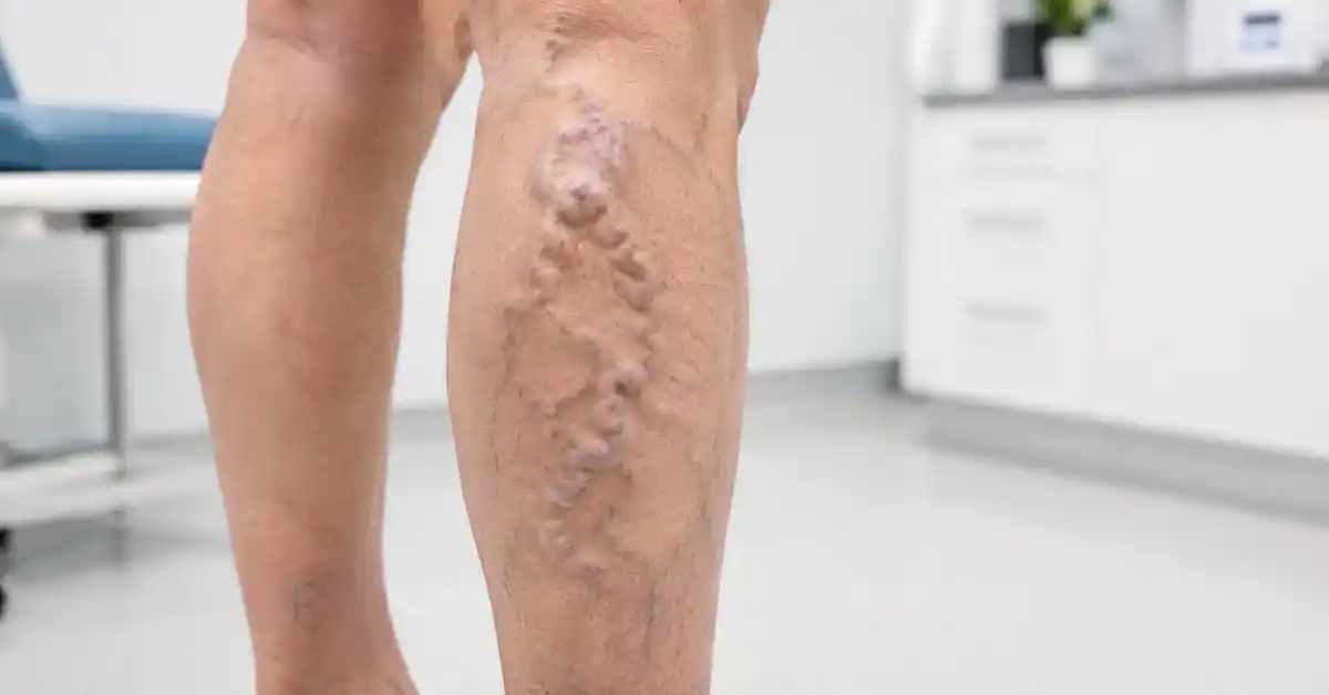
What Varicose Veins Look Like
Varicose veins are dilated, usually tortuous superficial veins measuring approximately 3 mm or more when assessed with the patient standing.
They can appear as:
- Blue or purple veins.
- Skin-colored enlarged veins.
- Twisted cords.
- Bulging vessels that can often be felt beneath the skin.
They are generally classified as clinical class C2 venous disease.
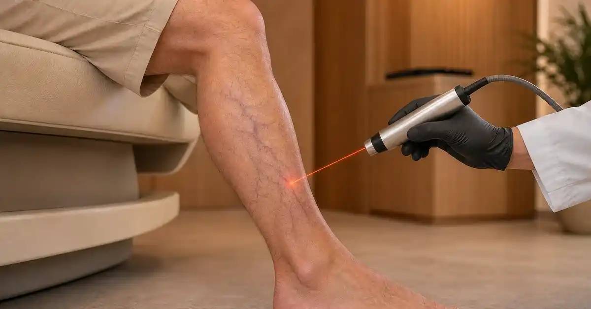
What Spider Veins Look Like
Spider veins, or telangiectasias, are very small superficial vessels usually measuring less than 1 mm.
They typically appear as:
- Thin red lines.
- Small blue or purple vessels.
- Branching or web-like patterns.
- Flat vessels close to the surface of the skin.
Spider veins and reticular veins fall within clinical class C1.
Reticular Veins Between Spider and Varicose Veins
Reticular veins are intermediate vessels measuring approximately 1 to less than 3 mm.
| Vein type | Approximate diameter | Typical appearance |
|---|---|---|
| Spider veins | Less than 1 mm | Fine red or blue networks |
| Reticular veins | 1 to <3 mm | Flat blue or green veins |
| Varicose veins | 3 mm or more | Bulging, enlarged, twisted veins |
Symptoms of Varicose Veins Compared With Spider Veins
The difference is not purely cosmetic. Larger varicose veins are more likely to be associated with symptoms of chronic venous disease.
Common Symptoms of Varicose Veins
Possible symptoms include:
- Aching.
- Leg heaviness.
- Burning or throbbing.
- Muscle cramping.
- Lower-leg or ankle swelling.
- Itching.
- Symptoms that worsen after prolonged standing or sitting.
Can Spider Veins Cause Pain?
Many spider veins are asymptomatic and mainly create cosmetic concern.
Some patients, however, experience localized burning, itching, or discomfort, particularly when there are extensive telangiectasias or reticular veins.
Severe symptoms or bleeding can justify evaluation for associated venous reflux.
Why Standing Can Worsen Venous Symptoms
Leg veins must move blood upward against gravity.
When valves do not close effectively, blood can flow backward and accumulate within superficial veins, increasing venous pressure.
This can make symptoms more noticeable after prolonged sitting or standing.
Causes and Risk Factors for Varicose and Spider Veins
The two conditions share some risk factors, although spider veins do not automatically indicate significant underlying venous failure.
Venous Valve Dysfunction and Varicose Veins
Varicose veins can develop when venous valves become incompetent and allow blood to reflux.
Risk factors can include:
- Family predisposition.
- Increasing age.
- Pregnancy.
- Prolonged standing.
- Prolonged sitting.
- Excess body weight.
- Reduced physical activity.
- Previous venous disease in selected patients.
Why Spider Veins Develop
Spider veins represent dilation of very small vessels within the skin.
They can be associated with genetics, age, hormonal factors, pregnancy, and prolonged standing, but they may also occur without significant reflux in larger superficial veins.
Routine ultrasound is therefore not recommended simply because asymptomatic spider veins are visible.
Can Spider and Varicose Veins Occur Together?
Yes.
A leg can have small spider veins, intermediate reticular veins, and larger varicose veins at the same time.
When spider veins occur alongside heaviness, swelling, pain, or obvious larger varicosities, assessment should focus on the overall venous system rather than the small visible vessels alone.
Cosmetic Spider Veins Versus Chronic Venous Insufficiency
One of the most important distinctions is whether the visible veins are an isolated cosmetic finding or part of progressive venous disease.
When Spider Veins Are Mainly Cosmetic
Small superficial vessels are more likely to represent a cosmetic concern when they are:
- Flat.
- Painless.
- Not associated with swelling.
- Not accompanied by skin changes.
- Not associated with large varicose veins.
Routine duplex testing is generally unnecessary in asymptomatic C1 disease.
Signs That Suggest More Significant Venous Disease
Seek assessment when visible veins are accompanied by:
- Persistent aching.
- Leg heaviness.
- Ankle or leg swelling.
- Chronic itching.
- Skin pigmentation.
- Venous eczema.
- A hard, painful superficial vein.
- Bleeding.
- A healed or active leg ulcer.
Possible Complications of Larger Varicose Veins
More advanced venous disease can be associated with:
- Superficial vein inflammation.
- Bleeding.
- Chronic edema.
- Skin damage.
- Venous ulcers.
- Thrombotic complications in selected situations.

Diagnosing Varicose and Spider Veins With Duplex Ultrasound
Many cases can be distinguished visually, but appearance alone cannot always determine whether deeper venous reflux is present.
Clinical Examination of Leg Veins
Assessment generally includes examination while standing.
The clinician evaluates:
- Vein distribution.
- Diameter.
- Bulging and tortuosity.
- Swelling.
- Skin changes.
- Tender or hardened veins.
- Ulceration.
Symptoms and their relationship to standing or activity also provide useful information.
When Duplex Ultrasound Is Needed
Duplex ultrasound evaluates blood flow and venous valve function.
It can identify:
- Superficial venous reflux.
- Deep venous abnormalities.
- The source feeding visible varicosities.
- The pattern that should guide treatment.
It is particularly important for symptomatic varicose veins and before treatment of significant underlying reflux.
Do All Spider Veins Need Ultrasound?
No.
Asymptomatic spider or reticular veins do not routinely require duplex evaluation.
Ultrasound can become appropriate when spider veins are associated with severe pain, burning, bleeding, or other signs suggesting deeper venous incompetence.
Treatment Differences Between Varicose and Spider Veins
The visible size of the vein and the underlying source of reflux determine treatment.
Treatment for Spider Veins
Options can include:
- Sclerotherapy.
- Transcutaneous laser treatment in selected vessels.
Sclerotherapy involves injecting a solution that closes the small vein so that it gradually becomes less visible.
More than one session may be needed when numerous vessels are present.
Treatment for Larger Varicose Veins
When duplex shows reflux in a major superficial vein, treatment may need to target that underlying source.
Options can include:
- Endovenous laser.
- Radiofrequency ablation.
- Selected nonthermal closure techniques.
- Foam sclerotherapy.
- Removal of visible tributaries through small punctures.
Why Treating Only Spider Veins May Not Be Enough
If visible small vessels coexist with significant underlying reflux, treating only the surface appearance may leave the main venous problem untreated.
The correct strategy therefore differs substantially between isolated cosmetic spider veins and symptomatic varicose disease.
Conclusion
Spider veins are usually less than 1 mm in diameter and appear as fine flat red, blue, or purple networks, while true varicose veins are generally 3 mm or larger and appear enlarged, twisted, and often raised above the skin.
Isolated spider veins are frequently a cosmetic concern and do not automatically indicate serious venous insufficiency. Larger symptomatic varicose veins, particularly when associated with swelling, heaviness, skin changes, or bleeding, require more detailed assessment to identify the underlying venous problem.
Frequently Asked Questions: Varicose Veins vs Spider Veins
Which is more serious, varicose veins or spider veins?
Larger varicose veins are more likely to be associated with venous insufficiency, symptoms, and complications. Isolated asymptomatic spider veins are often cosmetic.
Do spider veins eventually become varicose veins?
Not necessarily. They can share risk factors, but having spider veins does not mean larger varicose veins will inevitably develop.
Do spider veins require duplex ultrasound?
Not routinely when they are asymptomatic. Duplex can be appropriate when significant pain, burning, bleeding, or other venous symptoms are present.
Can spider veins be treated with laser?
Yes. Transcutaneous laser can be appropriate for selected small vessels, while sclerotherapy is another common treatment.
When should varicose veins be medically evaluated?
Persistent pain, swelling, heaviness, skin changes, a hard painful vein, bleeding, or a leg ulcer should prompt assessment.